
As Dr. Abrams' practice grew, spurred by the increasing accuracy and thoroughness of his diagnoses, more and more instances arose of patients who wished to avail themselves of his method but who could not visit his office, either because they were too ill to make an office visit, or lived too far away. In the effort to be of service to these individuals, experiments were made with blood specimens taken from people who were ill, and placed in a metal cup (which Dr. Abrams termed a "dynamizer"), with connecting wires to the head and foot plates attached to the healthy subject (reagent) on whom the percussion diagnosis was performed. It was found that the blood carried all the radiations of the body from which it had been drawn, and could therefore be used as a substitute for the presence of the patient in the examining room. With the blood in the metal cup connected to the healthy subject, the diagnostic tests performed on that subject produced the same tunings and measurements as those prevailing when the patient (from whom the blood had been drawn) was personally connected to the reagent, through the tuning controls.
Since the blood flowed through every organ, gland and tissue of the body, it was reasonable to assume that all the radiations from those organs, glands and tissues would be absorbed by the blood including the radiations of any diseases present. This theory was later proven independently by the blood crystallizations method developed by the late Dr. Ehrenfried Pfieffer. He perfected a technique for evaporating a small amount of blood in a copper sulphate solution. Crystalline patterns were formed, each different disease resulting in a different pattern.
The blood came to be used in the E.R.A. method (and later in radionics by some of the practitioners), in dried form, on a piece of blotting paper or other absorbent paper; this is in contrast to laboratory procedures, for which the blood is used, in liquid form. Since liquid blood spoils rapidly, it has to be mixed with a quantity of preservative. For the purpose of E.R.A. or radionic tests, a preservative must not be used, since it adds a strong foreign vibration or radiation of its own. Since dried blood serves equally well as liquid blood for the purpose of E.R.A. or radionic tests, the blood came to be used in dried form to avoid the use of preservatives.
The use of blood specimens broadened the utility of E.R.A. method but introduced special problems. One of the problems was that of contamination. Both the E.R.A. and the radionic method are extremely sensitive, to the extent that if someone other than the patient handled the piece of paper on which the patient's blood specimen was placed, even a very slight [14] amount of skin perspiration from the other person would place that person's radiations into the paper, to a degree. The result was that the practitioner would then likely detect the combined radiations of the two individuals -- the patient, and the Doctor or other person who had handled the specimens. This of course led to detecting and reporting of ailments or conditions not present in the patient. When this was discovered, special precautions were laid down for the preparation of blood specimens, to keep them free from contamination. However, those who would try to trick the practitioner were not interested in observing such precautions.
Another disadvantage of the use of blood specimens lay in the fact that the door was opened to trickery of various kinds. For example, blood specimens from animals and poultry were sent to the Doctor, ostensibly as human blood specimens. When the reports came back listing the various diseases found, the method was derided and ridiculed, Actually, the same trick can be successfully perpetrated on medical laboratories, since the laboratory work required to perform blood counts does not disclose whether the specimen is from a human or from an animal! The blood count ranges differ somewhat, but only to the extent that a reading that would be normal for some of the animals occurs frequently in humans who are ill. Animals and poultry are subject to the same toxic and infectious diseases as are humans; primates have the same organs and glands as do humans, quadrupeds have most of the organs, glands and other structures of humans. Poultry are now being used as the subject of spin tests in the astronaut program "because their circulatory system is so similar to that of humans". The reports of disease radiations found in the animal specimens did not in the least invalidate the method, but those unskilled in a new field are quite prone to unthinkingly accept adverse criticism that seems logical on the surface.
In modern radionics there are tuning rates to differentiate human and animal specimens; however this requires extra checking and so are not always used.
EVOLUTION OF THE DETECTION METHOD
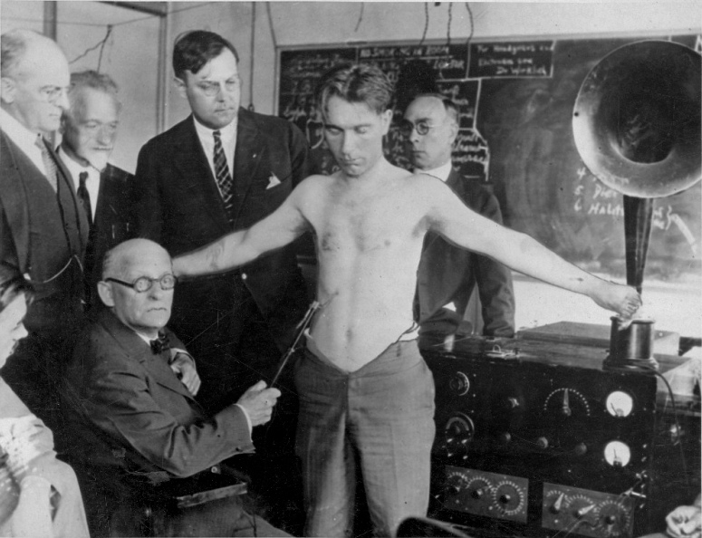
In the evolvement of the Abrams method of detecting disease radiations, the next step taken was the abandonment of the percussion tests, and in their place the adoption of the method of rubbing or stroking the abdomen of the healthy subject, with a glass rod. The percussion method of eliciting the signals or responses required a very high degree of skill, and was too difficult to teach to others. The use of the glass rod, while not easy to learn, at least proved possible for some others besides Dr. Abrams. In the use of the rod, the ease with which it slipped over the abdominal surface was noted. A significant manifestation occurred when the rod appeared to "stick" over a particular abdominal area, where more energy or force was required to move it a given distance on the skin of the subject. This phenomenon of "sticking" occurred in conjunction with tunings for disease and would disappear when the volume measurement control was advanced to a setting indicative [15] of a strength of radiation greater than that which was being received.
COLORED LIGHT AND DISEASE
A further development was the addition of colored lights to shine on some element of the detecting circuit. These were said to have been originally suggested to Dr. Abrams by a Dr. McManus, an osteopath from Kirksville, Mo. The original colors used were red, yellow, green and blue, in addition to incandescent light of nonspecific color, termed "white light". A selector switch was incorporated, permitting the use of any one of those lights, or the use of no light. The reason for the use of colored light was because it was found that certain disease radiations manifested more strongly in the presence of specific colors of light. Thus the TB radiation was stronger and more easily detected when the red light was used; staph. radiation was stronger when the blue light was on; strep. was more readily detected with green light; and inflammation came in more clearly when the yellow light was used. Bacteria, cold or flu toxins manifested better when white light was turned on.
(To be continued. Next instalment will include description of the E.R.A. treating equipment.)
* * *
MAGNETIC FIELD CANCER CURE
"Mice live up to 45% longer after they've been subjected to certain types of magnetic fields, and cancerous mice lose their malignant growths after similar treatment, a Los Angeles space scientist said here yesterday. (Los Angeles 'Times', April 1960). Dr Harold S. Alexander of North American Aviation Corp.'s missile division told scientists at an annual meeting of the Institute of Environmental Sciences at the Biltmore that much more research must be conducted before the effect of these magnetic fields on humans will be known.
"But, he said, several leading cancer researchers are already pursuing the experiments pioneered by Dr. Jeno Barnothy, Hungarian physicist now of Chicago, who participated in yesterday's program.
"'Aside from the effect on malignancies (cancer), we don't yet know why the mice live so much longer after four to six weeks in a magnetic field,' Dr. Alexander said, 'but we think the experiments have some effect on the rate of cellular reproduction.'
"He displayed photographs of two mice from the same litter which had reached an age equivalent to 90 years in humans. The one which had lived for awhile in a magnetic field appeared only about one third as old as the other."
The California Medical Association has now had over eight years to research this spectacular cancer-cure breakthrough, but so far only thunderous silence.
Continue with "The History and Development of Radionics" (Part V)
 was widely known in his own lifetime for the development of spondylotherapy, and his electricity therapy (Electronic Reactions of Abrams, ERA), which formed the basis for the later practice of radionics.")
Further Reading
- Abrams, Albert. New Concepts in Diagnosis and Treatment: Physico-clinical Medicine, the Practical Application of the Electronic Theory in the Interpretation and Treatment of Disease: with an Appendix on New Scientific Facts. San Francisco, Calif: Philopolis Press, 1916. Print. [Digital: <http://catalog.hathitrust.org/Record/001588073>; reprints are available through BSRF in our classic xerographic format: <#B0210, "New Concepts in Diagnosis and Treatment">]
- Abrams, Albert. Human Energy. San Francisco: Philopolis Press, 1914. Print. [Digital: <http://catalog.hathitrust.org/Record/008904901>; reprints are available through BSRF in our classic xerographic format: <#B0211, "Human Energy">]
- Abrams, Albert. Spondylotherapy: Physio and Pharmacotherapy and Diagnostic Methods Based on a Study of Clinical Physiology. San Francisco: Philopolis Press, 1914. Print. [Digital: <http://catalog.hathitrust.org/Record/002089090>; reprints are available through BSRF in our classic xerographic format: <#B0177, "Spondylotherapy">]
An introductory review of Abrams and his work can be found in
"ERA: Electronic Reactions of Abrams" (#B0025).